In The Diagram Which Labeled Structures Are Atrioventricular
Transverse section of human heart. left and right atrioventricular Circulatory the heart Valves circulatory chambers herzklappen blood herz klappen funktion visiblebody ähneln regulate
52 In the diagram which labeled structures are atrioventricular valves
Atrioventricular valves diagram Aortic valve Pulmonary valve
Pulmonary valve
What is a heart murmur? « fitzalan house veterinary surgeryPulmonary tricuspid mitral to better watch an animation anatomy de Solved drag the labels onto the diagram to identify the17.2b: operation of atrioventricular valves.
Tricuspid valve: overview, function and anatomyAtrioventricular valve function Atrioventricular valve diagramHeart anatomy.

How many hearts does a cow have
Blood flow through the heartPulmonary atria ventricles aorta cardiac veins vena superior inferior physiology Function of the atrioventricular and semilunar valvesBlood heart diagram flow labeled oxygenated lungs arteries major medicinebtg picture.
Semilunar valve definition and examplesLabeled diagram of the heart and blood flow Cardiovascular system anatomy and physiology: study guide for nursesLeft atrioventricular valve.

Valve valves atrioventricular pulmonary semilunar heart fibrous mitral skeleton function human aortic cusps tricuspid anatomy physiology outline diagram tissue between
Valve heart valves aortic pulmonary mitral cardiac surgery tricuspid anatomy valvuloplasty atrioventricular blood section left cross flow diagram human balloon52 in the diagram which labeled structures are atrioventricular valves Heart diagram: anatomy of the heart and its major vesselsHeart valves diagram labeled.
Images of av端子Valves atrium semilunar ventricle flap valve chambers tissue prevents flowing connective Valves valve anatomy cardiac nursing aortic pulmonary cardiovascular disease mitral tricuspid physiology aorta medicinebtg cardio leaflets valva atrioventricular murmur valvularWhat is the flap of connective tissue between an atrium and a ventricle.
:max_bytes(150000):strip_icc()/human-heart-circulatory-system-598167278-5c48d4d2c9e77c0001a577d4.jpg)
Collection 92+ pictures how do the highlighted valve cusps function
Lab 3, exercise 3 flashcardsCardiovascular physiology valves herz cardiac nurseslabs chambers aortic menschliche anatomie menschliches nurses cardio physiologie corazon maqueta body vessels circulatory anatomia The atrioventricular valve's are found between the atria and theHeart figure valve left right blood vessels ppt semilunar pulmonary veins aorta artery powerpoint presentation atrium.
Cardiac surg textbook scaredSemilunar biology Heart valves: types, structure, functions, diseasesHeart valves diagram labeled.

Heart blood myocardium vessels valves biology muscle figure bio layer atrium ventricle left right through valve vena cava into chambers
Solved 1. in the diagram, which labeled structure preventsValves heart valve tricuspid adult cardiac embryology papillary file location muscles tendineae located structure present histology definition function other intermediate Valve atrioventricular heart tricuspid valves backflow found two mitral between ventricles atria pulmonary medical prevent they choose boardHeart anatomy valves figure valve anterior physiology right ii four section mitral transverse tricuspid cusps left atria vessels atrioventricular different.
Valve aortic wikipedia heart diagram human wikimedia wiki upload cropped svgValves cow semilunar circulatory Mammalian heart and blood vessels.
.svg/1200px-Diagram_of_the_human_heart_(cropped).svg.png)

PPT - Heart and Blood Vessels PowerPoint Presentation, free download

Transverse Section of Human Heart. Left and Right Atrioventricular

Atrioventricular Valve Diagram

Tricuspid Valve: Overview, Function and Anatomy
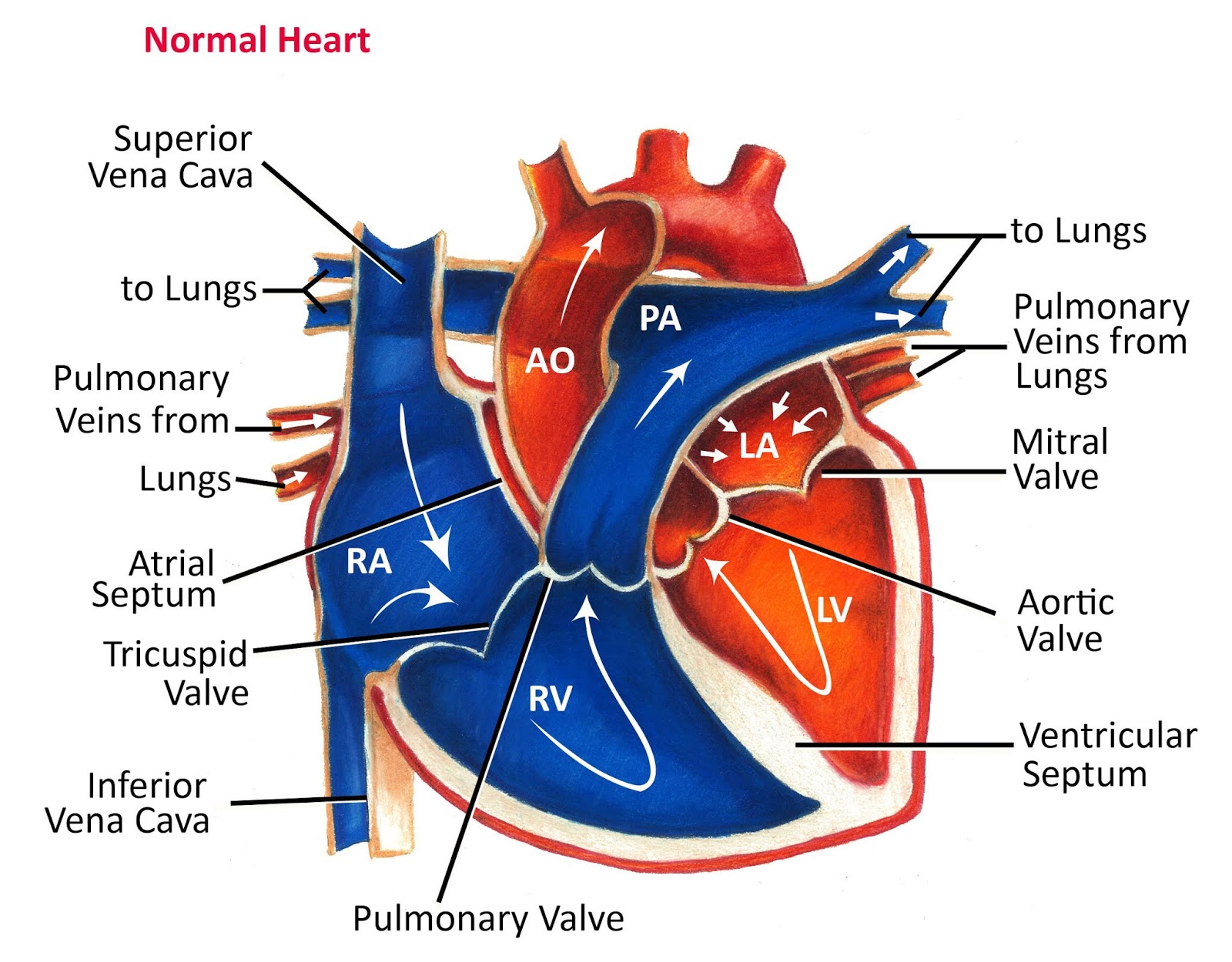
Blood Flow Through The Heart

Heart Diagram: Anatomy of the Heart and its Major Vessels

Atrioventricular Valves Diagram | Quizlet